Aïe! 40+ Raisons pour Shoulder Muscles Diagram Anterior! Posterior part of the deltoid:
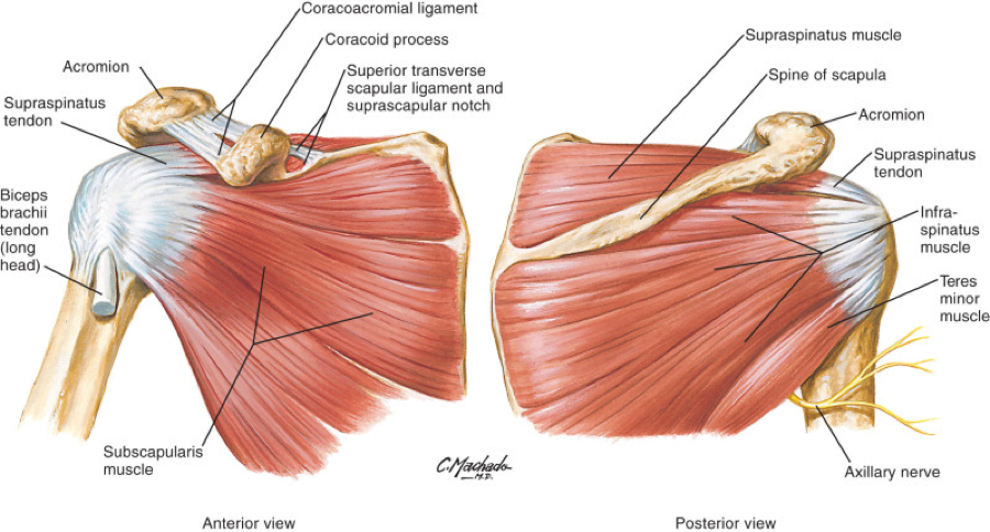
Shoulder Muscles Diagram Anterior | The anterior, lateral and posterior deltoid heads. The shoulder has about eight muscles that attach to the scapula, humerus, and clavicle. Shoulder stretching exercises, including anterior shoulder stretch, chest stretch, triceps… It keeps the scapula in position close to the chest wall, abducts the. Anterior surface of the medial clavicle. Anterior superior iliac spine insertion: The next life study seated female figure, shows the upper part of the the muscles of the superficial layer of the back move the shoulder blade (scapula) and upper arm (humerus). Right anterior basal segmental bronchus. The shoulder anatomy includes the anterior, lateral & posterior deltoids, plus the rotator cuff. Diagram demonstrating the sagittal plane of the external obliques. The thoracic muscles attach on the anterior and lateral regions of the thorax, or rib cage. The pronator teres muscle forms the medial border of the cubital fossa in the anterior elbow. Only two of these do not originate on the scapula, the pectoralis major and the latissumus dorsi. Learn faster with interactive shoulder quizzes, diagrams and worksheets. Produce wrist and/or finger flexion. The articulations between the bones of the shoulder make up the shoulder joints. They are shown in the image below. Extends and laterally rotates the arm. The shoulder has about eight muscles that attach to the scapula, humerus, and clavicle. The external abdominal oblique muscle is the largest and most superficial of the four muscles. Want to learn more about it? Muscles of the anterior compartment of the forearm. It is a functionally important muscle that contains two heads. Diagram shoulder muscles anatomy 101 shoulder muscles the handcare blog. Anterior surface of the medial clavicle. They're all small, simple muscles. These muscles form the outer shape of the shoulder and underarm. The muscles located in the anterior compartment are involved in flexion at the elbow and shoulder joint whereas muscle in the posterior compartment, triceps brachii the muscles of the anterior compartment are further divided into a superficial, intermediate and deep layer; Anterior and posterior shoulder muscles. The shoulder joint has the most range of motion of any joint on the. Produce wrist and/or finger flexion. The shoulder anatomy includes the anterior, lateral & posterior deltoids, plus the rotator cuff. The shoulder muscles are associated with movements of the upper limb. The anterior, lateral and posterior deltoid heads. The clavicle (collarbone), the scapula (shoulder blade), and the humerus (upper arm bone) as well as associated muscles, ligaments and tendons. This workout is great for your anterior deltoids, and it is often considered the most effective exercise for building. Sternum and superior six the pectoralis major muscle is the most important muscle for the adduction and anteversion of the. The anterior serratus pulls the scapula outward which lifts the shoulder. The system used here groups the muscles based on their function and topography (which are closely related in the upper limb) Rotator cuff is formed by a group of four muscles that surround the shoulder joint. Their main function is contractibility. Their main function is contractibility. The thoracic muscles attach on the anterior and lateral regions of the thorax, or rib cage. They are shown in the image below. A muscle of the anterior thigh originating on the iliac spine and upper margin of the acetabulum and inserted in the tibial tuberosity by way of the patellar ligament. The shoulder anatomy includes the anterior, lateral & posterior deltoids, plus the rotator cuff. Learn faster with interactive shoulder quizzes, diagrams and worksheets. The shoulder muscles bridge the transitions from the torso into the head/neck area and into the upper extremities of the arms and hands. Medial tibia near tibial tuberosity action: Anterior part of the deltoid: Flexes and medially rotates arm; Tutorials on the shoulder muscles (e.g rotator cuff muscles: Anterior surface of the medial clavicle. They're all small, simple muscles. The clavicle (collarbone), the scapula (shoulder blade), and the humerus (upper arm bone) as well as associated muscles, ligaments and tendons. Anterior to the interosseous membrane. Extends and laterally rotates the arm. The shoulder joint has the most range of motion of any joint on the. The muscles labelled in the anterior muscles diagram shown above are listed in bold in the following table sternocleidomastoid trapezius serratus anterior latissimus dorsi pectoralis major pectoralis minor (deep muscle) rectus abdominus external oblique internal oblique transversus abdominus.


The anterior deltoid, the lateral deltoid, and the posterior deltoid shoulder muscles diagram. Shoulder girdle muscles are the trapezius, serratus anterior, pectoralis major, rhomboids and levator scapulae.
Shoulder Muscles Diagram Anterior: Posterior part of the deltoid:
Refference: Shoulder Muscles Diagram Anterior
0 Tanggapan